Category: Uncategorized

Aug 01

X-ray image quiz

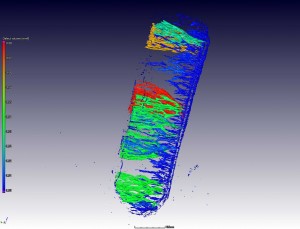
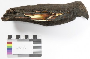
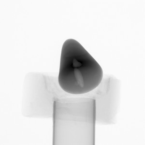
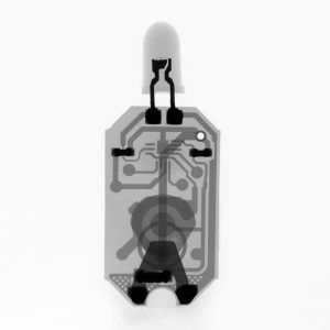
The quiz for March is this one, can you guess what is X-rayed here: The last solution was: a tensile specimen, in this case 5.5 mm diameter round gauge 20 mm in length, with Ti64 alloy with internal pores visible in the X-ray image. The latest X-ray image solution: a venomous snake fang. Why was …

Continue reading

May 31

Image based simulations: fluid flow in wood microstructure

This example showcases the capability of high resolution 3D images combined with simulations. A nanoCT scan of wood microstructure was used as basis for a simulation of fluid flow, taking as input parameters water viscosity, sealed edges and a pressure difference from top to bottom through the wood. The simulation then provides visual and quantitative …

Continue reading

May 31

Reverse engineering of a shoe

This demonstration shows how a shoe can be scanned and internal dimensions extracted in the form of a STL file, suitable for CAD programs. Engineers could use this to test their designs and to compare how a new and worn shoe differ, or how different shoes differ in internal geometry, which is where it counts. …

Continue reading

Nov 19

Head of the CT Lab

In this example, an indigenous South African chameleon species was scanned, which has three horns on its head making it quite unique. This example will be published as a part of a later scientific article but we show some selected images here as a sneak preview.   A closeup scan of the head was done …

Continue reading

Nov 19

Nondestructive testing of welded steel cylinder

X-ray testing of metal parts is used widely to find flaws, defects, cracks, pores or even inclusions which all can have serious effects on the part’s mechanical properties. CT scanning provides an “X-rays on steroids” advantage. Much more detail is visible and full 3D data can be analyzed to create a colour coded thickness analysis …

Continue reading

Nov 19

Ostrich shell microstructure

Ostrich eggs are very strong, yet ostrich chicks are able to peck their way out from the inside with little force. Investigating the inside of a fragment of ostrich shell might shed some light on this interesting natural mechanical system. These preliminary results show a fragment scanned using the nanoCT scanner and analysed for porosity …

Continue reading

Sep 23

A view into Ancient Egypt through a mummy falcon

An ancient Egpytian falcon mummy was analyzed at high resolution in a “virtual autopsy” and it was found that it’s stomach contains a mass of bones and teeth of two or more mice as well as a small bird. This leads us to believe the ancient Egyptians kept birds of prey in captivity, since a …

Continue reading

Sep 23

Drill core analysis

Geological drill core analysis is useful for exploration purposes, and when you understand your sample CT data can provide quantitative ore grade information as shown in this case study, published in Computers & Geosciences. The results correlate well with industry standard methods, with a hugely improved turnaround time compared to traditional analysis laboratories plus additional …

Continue reading

Sep 23

X-ray competition

We are having an X-ray image competition, and the winner gets a full 3D scan and 3D print of an object of his choice (plus free publicity). Additional sponsored prizes are planned but not yet confirmed. The two categories are as follows: Weirdest object X-ray’ed Most striking X-ray image How it works: you contact our …

Continue reading

Jul 31

Ghent nose CT scan

A Ghent nose is a sweet originating from Ghent, Belgium. Since we visited Ghent last month, we decided to scan and analyze a Ghent nose as an example. This example shows how 3D X-ray imaging can be used to visualize internal features of food products, in this case showing clearly some large air bubbles / …

Continue reading