The Hans Merensky Chair in Measuring and Modelling Eucalypt Growth & Wood Formation
Understanding growth in the world's most widely planted hardwoods
Renewed use of newly upgraded microscope in capturing high quality images
Post authored by Lucy Nevhungwili
The concept of performing good image analyses lies in capturing high quality images.
To enable one to use image analysis software, such as ImageJ and QuPath, to identify and quantify different cell types, crisp and clear images are needed. Last month, we finally upgraded the Nikon Eclipse E400 microscope with a Prior XYZ stage and installed the NIS elements software on the controlling computer. These upgrades allow one to stitch images taken of large samples with the XY functionality, while the Z movement allows one to stack images easily, which results in images that are in focus right through, while eliminating over exposure (Figure 1).
(a) 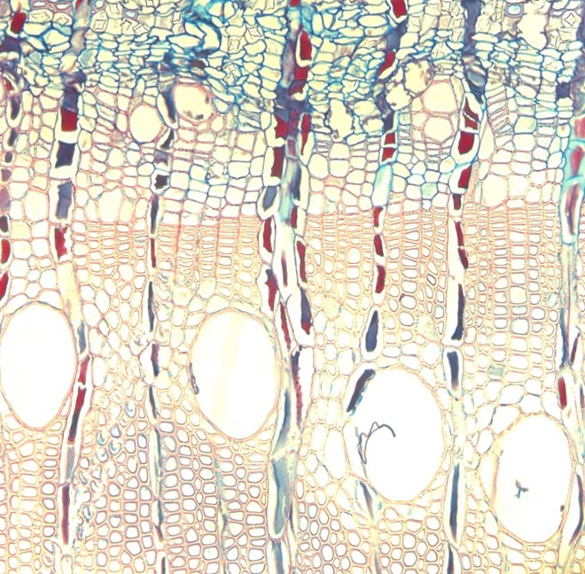 (b)
(b) 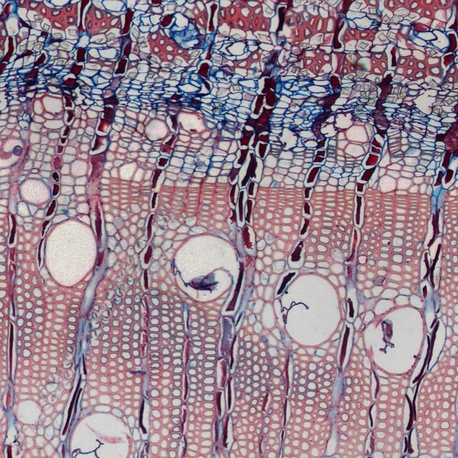
Figure 1. (a) Image of a Eucalyptus microcore taken before the upgrade and (b) taken with the newly upgraded microscope.
This upgrade was of huge help to one of our master’s students, Miss Lucy Nevhungwili, who is currently working on visualising and quantifying the size of the vascular cambium in Eucalyptus. This tissue is characterised by extremely thin cells that are very close to one another, and it is often difficult to obtain high quality images of it because the cambium tends to distort during sectioning.
Below is one of the challenges that Lucy was facing before the upgrade, but now using the newly upgraded microscope, she can use the stitching and stacking functions to create images of complete wood cores (Figure 2), which increases the area to select the region of interest from.
 Figure 2. Image of a complete Eucalyptus core taken at 100x magnification.
The team is looking forward to doing a lot more imaging on the newly upgraded Nikon Eclipse E400 microscope!