A look back at 2019: Staff training on Super-resolution microscopy in Scotland

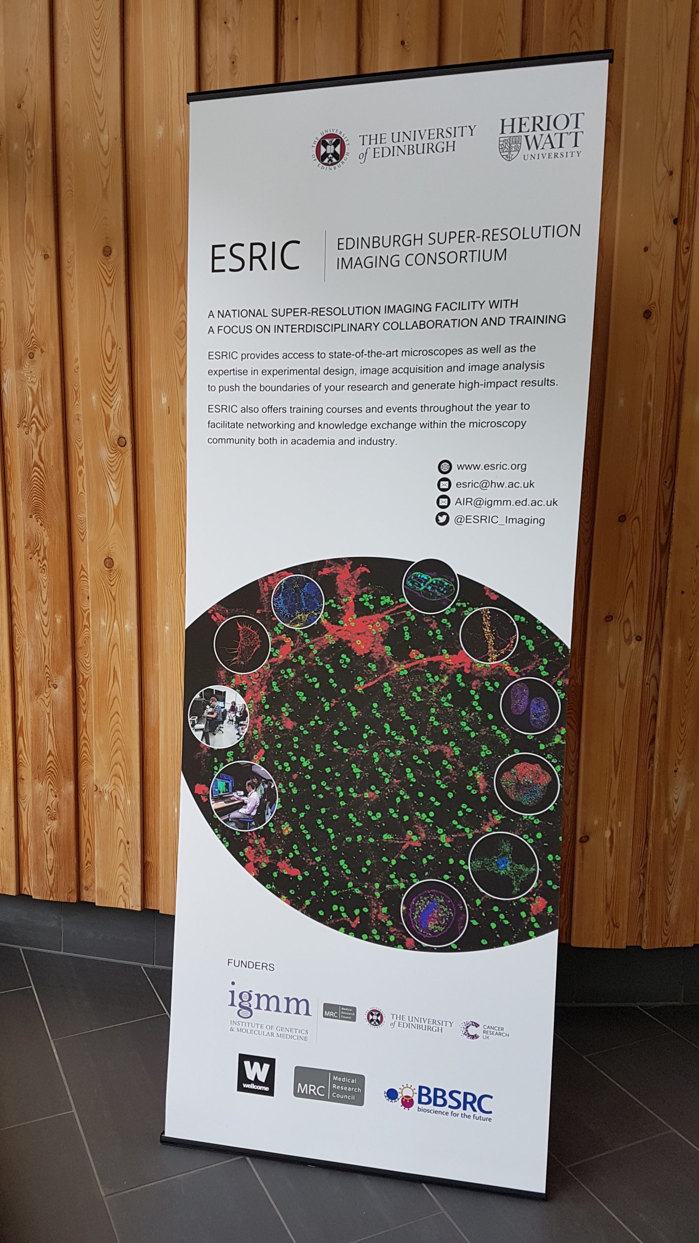
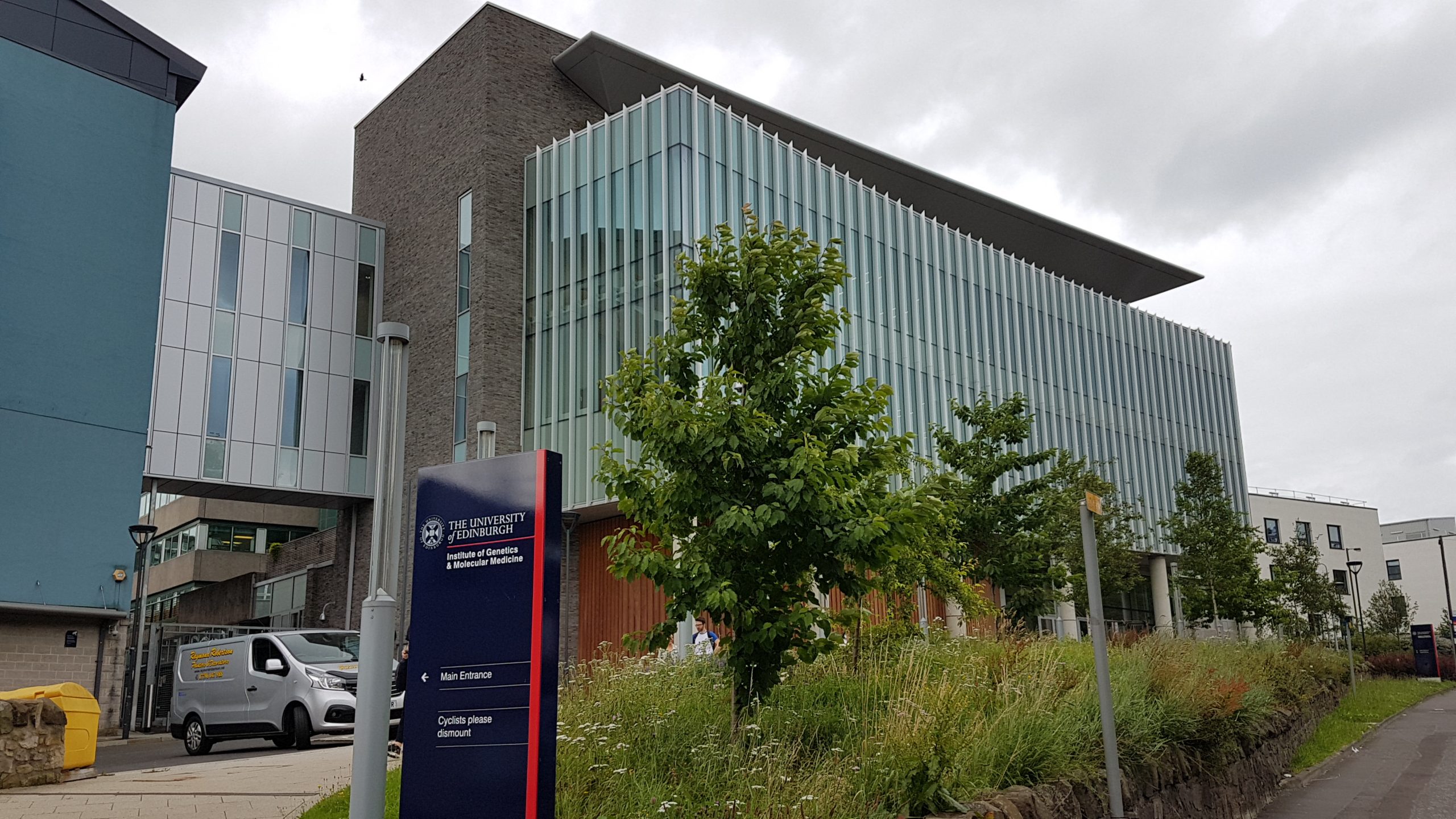
Lize Engelbrecht, manager of the Fluorescence Microscopy Unit was the first South African selected to attend the ESRIC super-resolution microscopy Summer School 2019 in Edinburgh. The Edinburgh Super-Resolution Imaging Consortium (ESRIC) is a National Super-resolution Imaging Facility with a focus on interdisciplinary collaboration and training. It is a joining of expertise across a broad scientific spectrum from cell biologists to physical scientists.

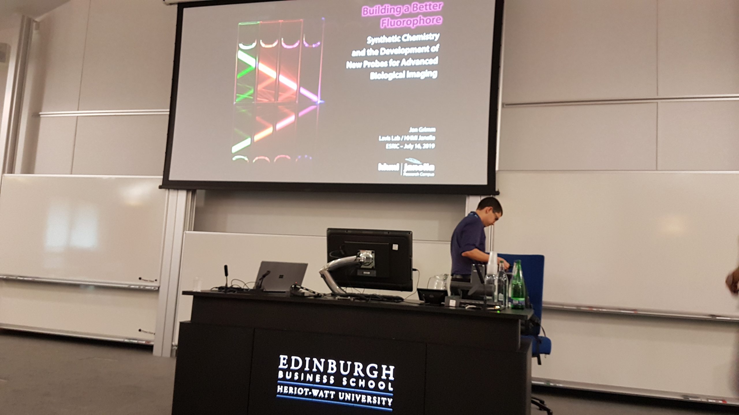
The ESRIC summer school is an annual advanced imaging training course which takes place over 5 days in Edinburgh. In 2019 the 6th Summer School were held, where only 36 delegates (3x as many applications) are selected to attend from all over the world. Delegates included researchers from the level of MSc and PhD up to professors, all who were interested in the techniques of super-resolution microscopy, how best to apply them to their research question, to meet fellow scientists in the super-resolution community and exchange knowledge and ideas. The work of all aimed to extend the boundaries of cellular imaging beyond diffraction limits in order to investigate cellular functions and human disease.

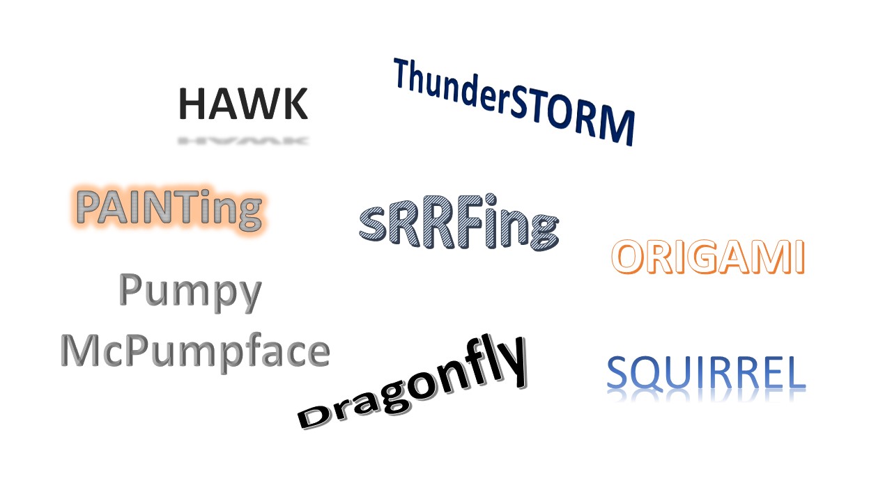
Topics included new developments in Structured Illumination Microscopy, STED and Single Molecule Localisation Microscopy (SMLM) and a large number of new acronyms and buzz words could often be heard during the workshop. Although most of these sounded like ordinary words, they actually referred to new plugins for current imaging processing software, new techniques of sample preparation and improved labelling techniques, as well as ways to validate and cross check super-resolution data.

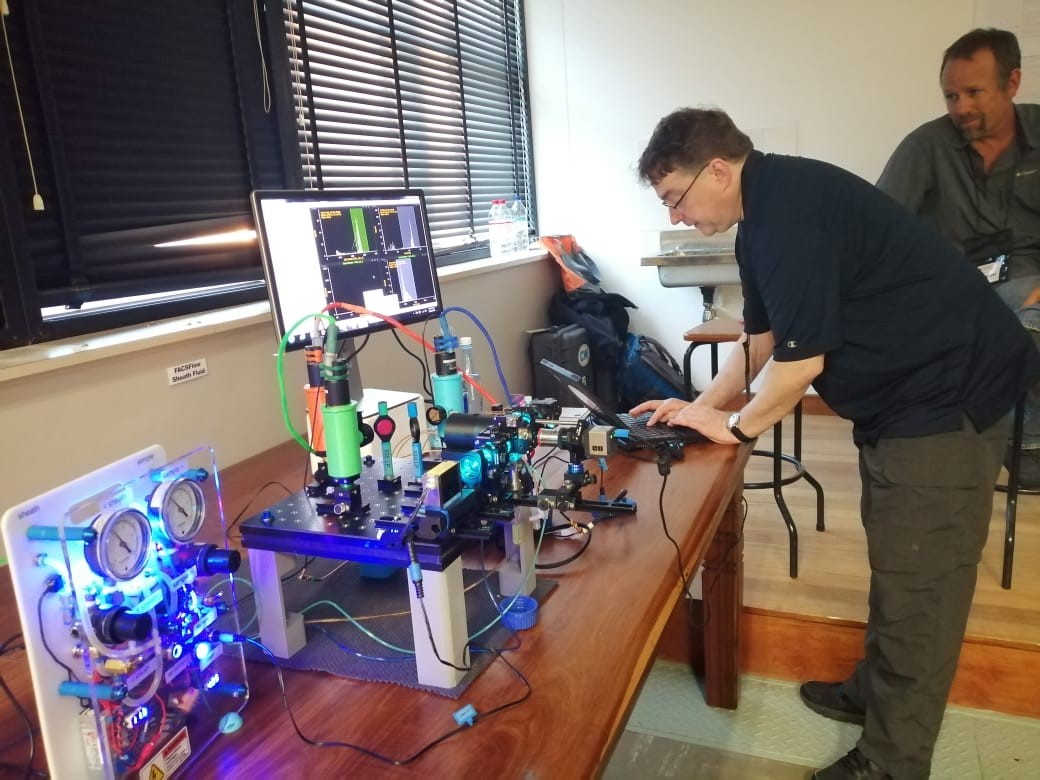
This was a very valuable opportunity for CAF to gain more experience and expertise in the field of super-resolution microscopy. We are looking forward to apply these techniques in many of the research projects here in South Africa.

A look back at 2019: Arrival of the new AMNIS ImageStreamX

Stellenbosch University was awarded a NRF National Equipment Programme Grant to purchase a new imaging flow cytometer which would be housed at Tygerberg Medical Campus.  The instrument arrived in June 2019 and CAF staff, along with the key applicants, prof Samantha Sampson and prof Carine Smith, along with their research group members received intensive training for three days at the beginning of July. Soon after, the research samples were run on the equipment and some great results have already been acquired.

The ImageStream®X MKII 7 is fitted with 7 lasers and 12 imaging channels. It delivers multiple images of every cell in flow, including brightfield, darkfield (SSC) and up to 10 fluorescent markers at high speed. The camera operates with a pixel size of 0.1/0.25/1 μm2 with 60X/40X/20X magnification respectively, allowing visualization of fluorescence location from the membrane, cytoplasm, subcellular organelles or nucleus at high resolution.

The innovative design of Amnis® cytometers increases signal and minimizes noise to provide exceptional photonic sensitivity. Design details like a dedicated side scatter laser, adjustable laser intensities, and brightfield imagery for the direct measurement of cell size allow the systems to resolve cell populations more effectively than far more expensive cytometers. The ease of use, outstanding performance, and imagery of each cell meet the needs of flow cytometry novices and experts alike.

We believe that this new addition will take research in the medical field at Tygerberg to the next level.

Assistance is available and if you need more information on the equipment, please have a look at the instrument specifications here: AMNIS ImageStream®X MKII 7

A look back at 2019: The second ISAC Cape Town Flow Cytometry Workshop


From 28-30th of May 2019,  our unit hosted the 2nd ISAC Cape Town Flow Cytometry Workshop, in partnership with the International Society for the Advancement of Cytometry (ISAC) . The workshop was held at the Tygerberg Medical Campus.

Seven internationally acclaimed experts in flow cytometry and some of our local experts in Cape Town conducted lectures and wetlabs on a range of topics, including stem cell analysis, cell cycle analysis,  polychromatic flow cytometry, intracellular cytometry  and cell signaling , electrostatic sorting and measures of quality control. A highlight was the demonstration on the assembly of a flow cytometer from all its most essential components.

Tutorials were presented and equipped the delegates to plan flow cytometry experiments effectively, analyse their data correctly and apply the correct quality control strategies.

This workshop included approximately 50 participants from academia and industry all across South Africa, with some participants joining us from other countries within Southern Africa. Overall, this workshop was very well received, and sparked discussions between the ISAC faculty and participants regarding project planning, quality control, troubleshooting, etc.

Overall this workshop was a great success, with both the ISAC faculty and the participants thoroughly enjoying the experience. Our staff members worked hard to make this a world class experience. We hope to see you at the next ISAC course!


All delegates and faculty members at the ISAC Flow Cytometry Work-shop in Cape Town.

Dr Bill Telford (Center for Cancer Research National Cancer Institute MARYLAND, USA) presenting a demonstration on how to build your own flow cytometer.

Alan Saluk (Scripps Institute, USA) presenting a wetlab on electrostatic sorting.

Dr Krishnamurthy (Central Imaging and Flow Cytometry at the National Centre for Biological Sciences, Tata Institute of Fundamental Research, Bangalore, India) preparing samples during the wetlab on DNA cell cycle and ploidy analysis.