Computed Tomography, CT Scanning, X-ray tomography, X-ray microscopy… all terms to describe the limitless capabilities of this technology that we offer to you.
As a CT Scanning service provider, our aim is to provide high-quality 3D imaging and analysis services. We welcome clients from both academia and industry.
Background to CT
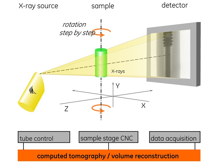
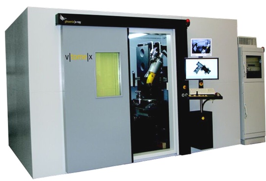
The Industrial Computed Tomography (CT) Scanner is an x-ray inspection machine that allows 2D x-ray inspections of materials as well as 3D CT Scans of materials, to investigate and analyse the inside of any object non-destructively and at high resolution and contrast. The measurement principle is similar to medical CAT scanners. An object is exposed to collimated x-rays and the absorbed radiation is measured with a sensor on the opposite side. This procedure is repeated from different angles around the object which allows a full 3D reconstruction. The technique is used to detect small differences in density and atomic number of the scanned sample. CT scanning provides state of the art non-destructive analysis of material properties for a wide variety of materials. Shown below are the machine itself, and a schematic illustrating the measurement principle.
What is it used for?
CT can be used to measure accurate distances, for example inside mechanical components, can be used to check porosity of materials, can be used to identify cracks, transitions or inclusions in materials, can be used to study the density variations within a sample, can be used to make a 3D CAD surface or volume data sets of the scanned object (eg. for reverse engineering). Standard 2D or 3D inspection reports are available, batch processing of large numbers of samples is possible, data can be provided in various formats such as jpg image slices, image stacks (this allows you to scroll through your object virtually in thin slices), dicom stacks (medical format), stl files (surfaces), or volume files (for self analysis of large volume data). Further analysis and high-end processing hardware and software is also available in our facility.
For students who use our facility, the generic experimental description is provided below with typical parameters (fill in your own parameters, details can be found in the PCA file with your data):
X-ray micro computed tomography (microCT) was used in this study. A General Electric Phoenix V|Tome|X L240 / NF180 was used. X-ray settings were 160 kV and 100 microA, 2000 images were acquired in a full rotation at image acquisition time of 500 ms per image, with no averaging and no skipping of images. Detector shift was activated to minimize ring artefacts. Background calibration was performed and the scan time was approximately 40 minutes per scan. Reconstruction was done with system-supplied Datos reconstruction software. Analysis was performed with Volume Graphics VGStudio Max 2.1 or Visualization Sciences Group Avizo Fire 8.0 commercial 3D analysis software packages.
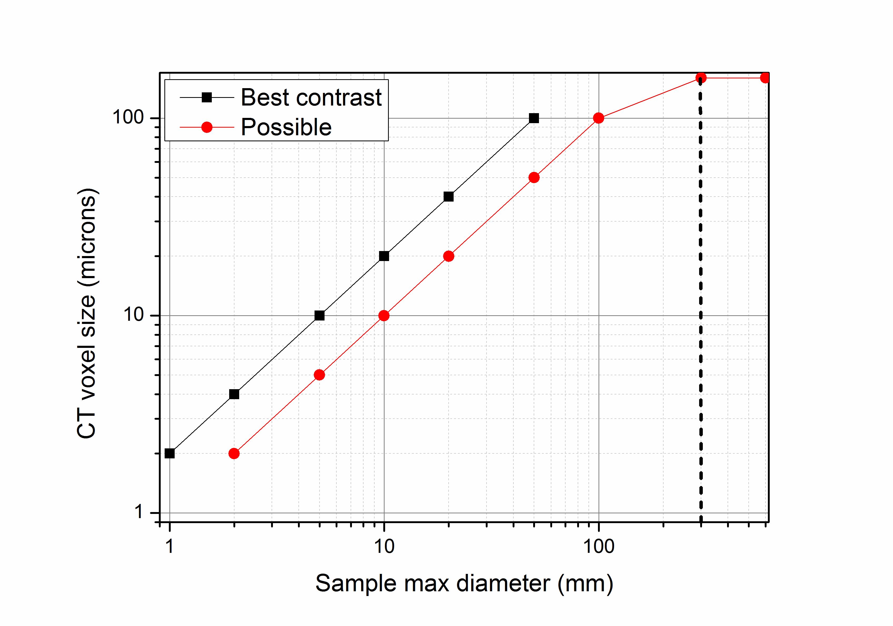
Sample size
CT System
General Electric Phoenix VTomeX L240 with NF180 additional x-ray source. This system is integrated with Phoenix Datos acquisition and reconstruction software.
CT data analysis
We extensively and almost exclusively use Volume Graphics VGSTUDIO MAX 2024.1.
For more information, please refer to their website.
 Follow