[:en]Imagine if the human body was as transparent as a jellyfish. This would mean that as we take a bite of an apple, we can track how it goes from the mouth to the stomach, how it gets digested and circulated all through the body. Life will be a lot easier for scientists if this were the case.
The bacterium that causes Tuberculosis– Mycobacterium tuberculosis, M.tb for short enters the body through inhalation. It then travels to the lungs where it stays and causes an infection. Looking at an infected patient, we cannot see the bug neither can we see how it causes an infection. We definitely cannot cut the patient open just to see what is happening inside. How then do TB researchers understand how TB causes disease? The answer is simple: we mimic and recreate.
We know that our body is made up of billions of cells and that each cell is functional on its own. Over the years researchers have been able to isolate living cells from humans and grow it up in a little dish, provided we give it the right nutrients and temperature conditions. How amazing! Similarly for bacterial cells like M. tb, we can isolate it from a TB patient, grow it in a dish and use it for different experiments.
The use of cellular models, such as this, is a widely appreciated technique in biomedical research. In TB research, it helps us answer questions such as-
- How does the bacterium cause an infection?
- How does the bacterium fight our human cells and survive without getting killed?
- What chemicals are capable of killing the bacteria?
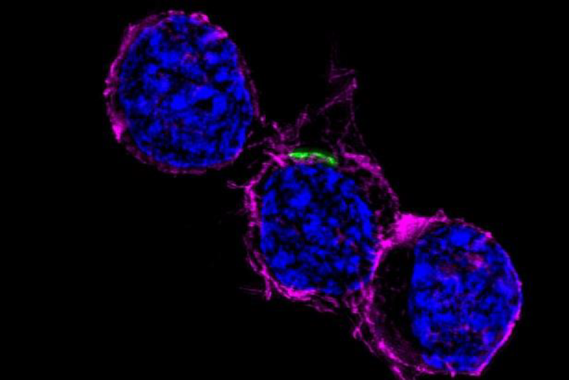
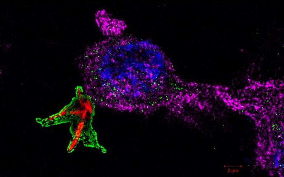
Another interesting technique in biomedical research is Fluorescence Microscopy. This involves the use of differentiate colors to label different cell components under the microscope. The cells, initially colorless can be made to fluoresce or emit a particular color of light by using external dyes. Whichever way, when viewed under a fluorescent microscope, different organelles or cells can be color coded.
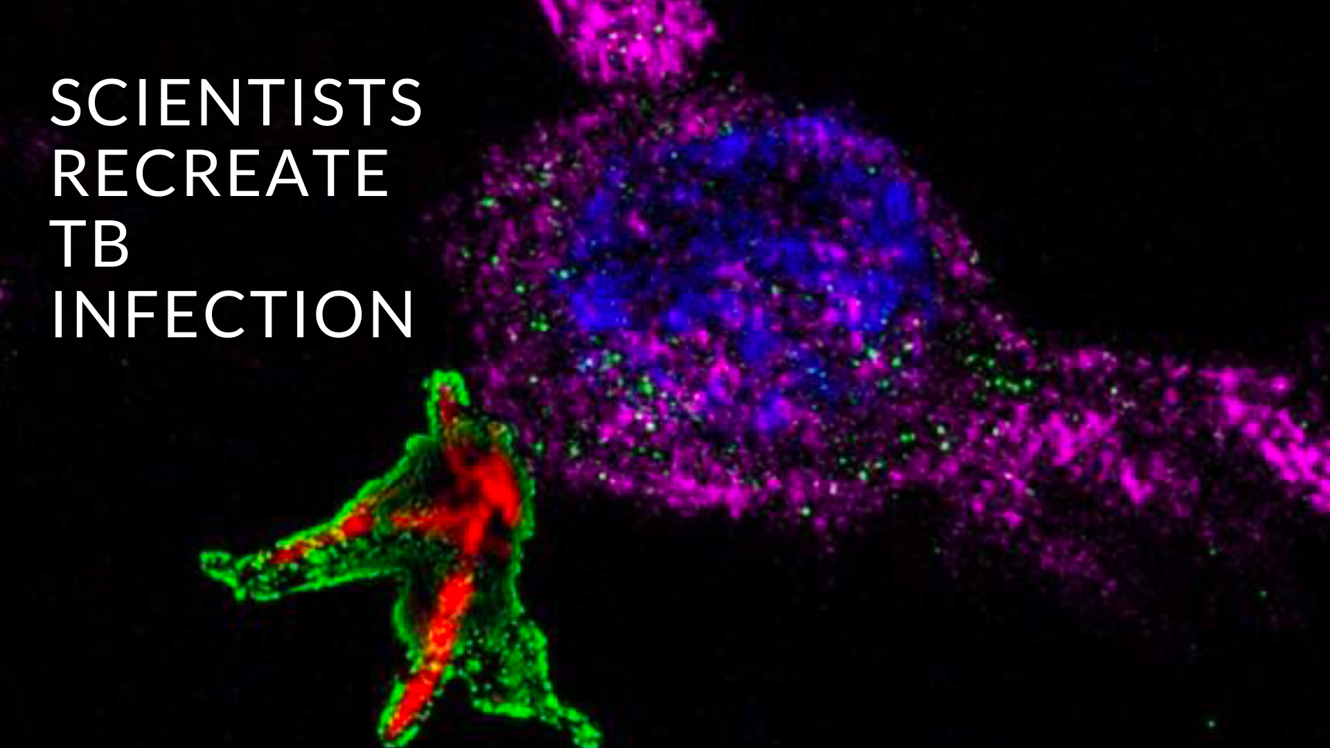
Combining cellular infection models and fluorescent microscopy, researchers can make bacteria fluoresce a particular color, use that to infect a cell and see what happens under the microscope. One can then investigate how the immune system gets rid of unwanted bacteria.
The human immune system is composed of many different types of cells. Phagocytes are cells that primarily function to swallow foreign invaders and degrade them inside the cell. They then display the digested components on their cell wall as a way of alerting and activating surrounding cells. Phagocytic cells include macrophages, dendritic cells and neutrophils. Alveolar macrophages are macrophages resident in the lungs and are the first cells to encounters of M. tb. Their first response is to engulf and restrain the bacterium in a phagosome. The phagosome is then prompted to fuse with an acidic lysosome forming a phagolysosome. Formation of a phagolysosome allows the degradation of contents by the acidic enzymes present in the lysosome. This is one of the ways a macrophage cell kills M. tb.


Researchers are currently using these techniques to investigate how M. tb is able to escape this phagosome and cause harm to the host. More observations like this help us to understand TB disease allowing us design better TB medicines. Now, think about the endless observations that can be made using cellular models and fluorescence microscopy!
 Written by: Ms Naomi Okugbeni
Postgraduate level: PhD (Molecular Biology) at Stellenbosch University node of the DST/NRF Centre of Excellence for Biomedical Tuberculosis Research housed within MBHG
Ms Okugbeni is currently a PhD candidate within the TB Host Genetics Research Group at MBHG. Her project aims to characterize the autophagic response to infection in M. tb-infected macrophages. Ms Okugbeni’s article was voted runner-up in the PhD category of the CBTBR Science Communication Awards.
[:]

