
This is a simple easy to understand example of what microCT scans can visualize. Everyone understands what a matchstick is composed of – a blob of dense material on top of a wooden stick. The full 3D data set shows inclusions (dense particles) inside the dense blob and the wood structure is visible (wood cells). The first image is a normal x-ray image, followed by full 3D CT images

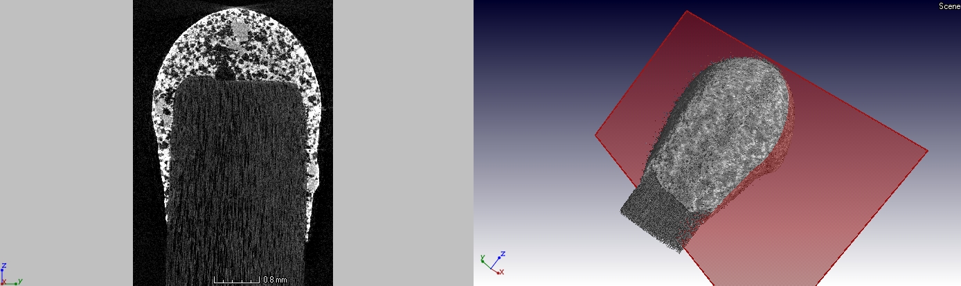
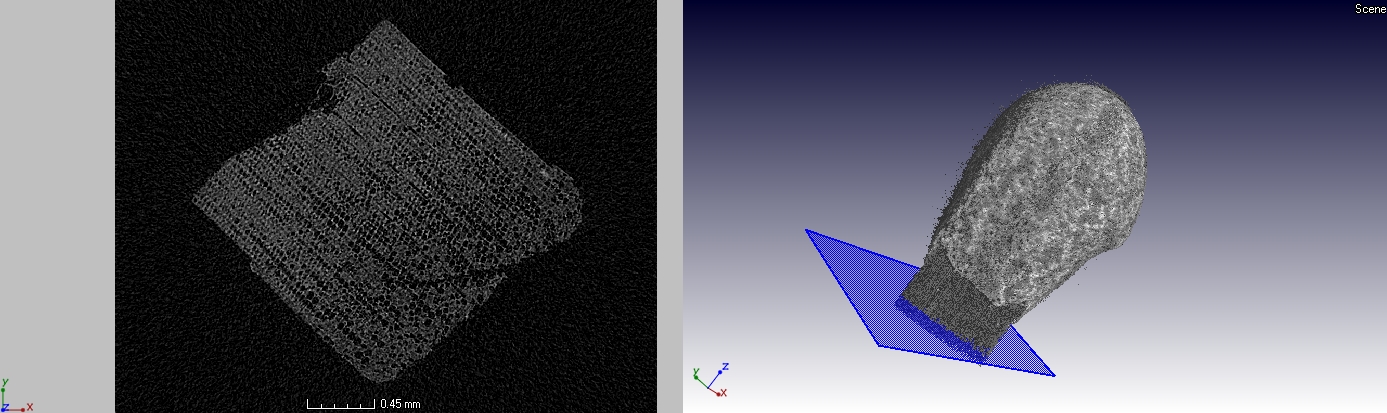
Note: this was a quick scan to demonstrate the principle only, higher quality and lower noise is possible with longer scan times
 Follow