This study in collaboration with Dr Karen Cloete from iThemba LABS includes some great coffee bean images and a great video can also be seen here. Find the full paper here: https://doi.org/10.1016/j.fochx.2019.100032 And a great video here: coffee bean v2
Category: Biological
 Follow
Mar 24
3D imaging of a wasp hive
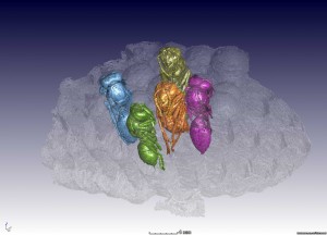
We scanned a wasp hive and found some interesting contents, which is a nice example of the non-destructive imaging capabilities of microCT. The 5 wasps are coloured for visual impression and all have been dead for a few months prior to the CT scan. Watch a 3D video of this example (link below the images). …
Apr 24
Maize kernel analysis
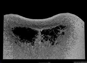
Maize is a staple food in South Africa and in Africa. One of the factors affecting the milling quality and efficiency of production is the maize hardness – in this study MicroCT was used to investigate hardness in maize kernels using full 3D information as well as a density calibration. Read the full paper here: …
Dec 04
3D Printing of samples scanned at the CT Scanner
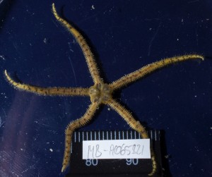
Recently we scanned another brittlestar (Brooding Ophiuroid). Due to the size of the sample (25mm) it was difficult to observe small features of the specimen. This granted us the opportunity to see how CT Scanning combined with 3D printing can enhance the way we can visualize and observe the small samples and specimens. In the …
Nov 20
Sample size vs resolution example
The most common misunderstanding people have the first time they use either microCT or nanoCT is how the sample size relates to the achievable resolution. In the following example I will try and provide you some insight into how these two parameters work together. To illustrate the example I scanned a packet of cigarettes at …
Oct 30
Biology – looking inside brittlestars
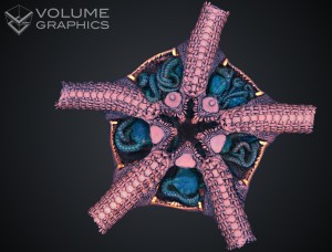
This work is from Jannes Landschoff, who won the best poster award for his poster “How many inside? A 3D Micro CT-Scan of Brooding Ophiuroids” at the European Echinoderms Colloquium, Portsmouth, UK in July. Jannes is a MSc student from UCT Biology. This work is still ongoing so more striking images will be loaded once …
Sep 26
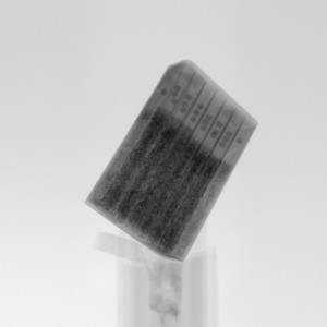
NanoCT of a toothpick
This example puts a nanoCT scan into perspective, as the 550 nm resolution allows a field of view of up to about 1 mm, allowing the tip of a toothpick for example to be analyzed in full 3D. This is why this is also called a 3D X-ray microscope. See below some images and videos …
Jul 31
Botany CT Scans
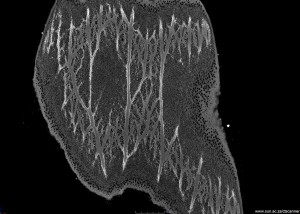
In this example, preliminary work was done by Prof Guy Midgley investigating the internal structures of selected South African plant leaves. One succulent and one Leucadendron laureolum leaf was scanned at different resolutions, though even higher resolution and detail is still possible. See below images of the succulent and images and a movie of the …
Jul 22
Consumer product testing: cookies

Non-destructive testing of consumer products is not new, with many laws protecting consumers. However, producers and suppliers do not test products unless explicity legally required. We offer simple analysis and testing for any users. In this example, we illustrate how a packet of cookies can be quickly inspected by CT scanning and X-ray imaging, with …
May 19
Abalone shell

This example is an abalone shell with coral on top of the shell. Some of the images show the internal connected pathways in the coral.