
The most common misunderstanding people have the first time they use either microCT or nanoCT is how the sample size relates to the achievable resolution. In the following example I will try and provide you some insight into how these two parameters work together. To illustrate the example I scanned a packet of cigarettes at a range of different resolutions in order to show how sample size influences this.
The main parameters of both CT scanner that must be kept in mind is 1) the detector used to capture the image and 2) the position of the sample relative to the X-ray source/tube.In order to capture the entire sample the sample image must fit in the field of view of the detector (Figure 1).
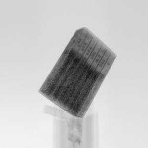
Figure 1: Indicates how a X-ray images of the sample looks when the entire sample is visible on the detector
The full pack of cigarettes was scanned at 100 micron resolution. In order to increase the resolution of the scan the samples is moves closer to the source/tube which decreases the field of view of the sample. To be able to do a 500 nano meter scan of a sample the sample must be small and almost on the X-ray source/tube (Figure 2). The distance between the X-ray source/tube a sample is approx between 4 and 1 mm.

Figure 2: Scan performed at 500 nano meter on a piece of tobacco from the cigarettes
In order to provide an idea of how the resolution, size of the sample and its location relative to the x-ray tube is related I combined a range of images depicting the scanning of the sample at 100, 50, 5 an 1 micron.

Figure 3: Pack scanned at 100 micron resolution
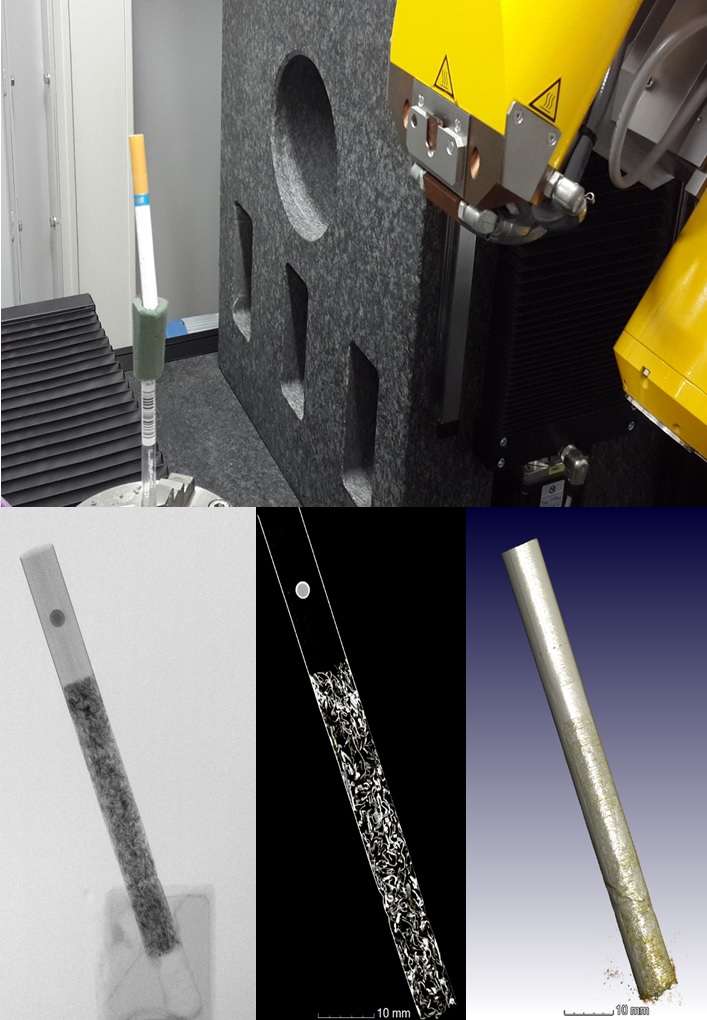
Figure 4: Single cigarette at 50 micron resolution

Figure 5: Single cigarette at 5 micron resolution
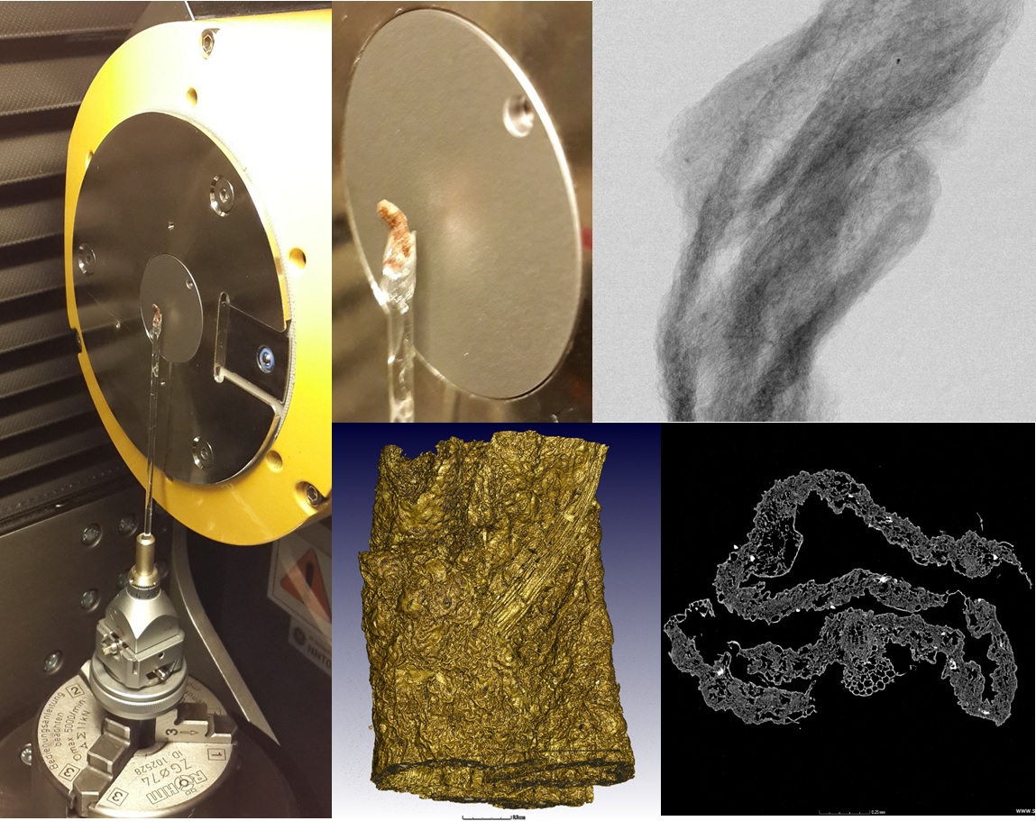
Figure 6: Piece of tobacco at 1 micron resolution
 Follow