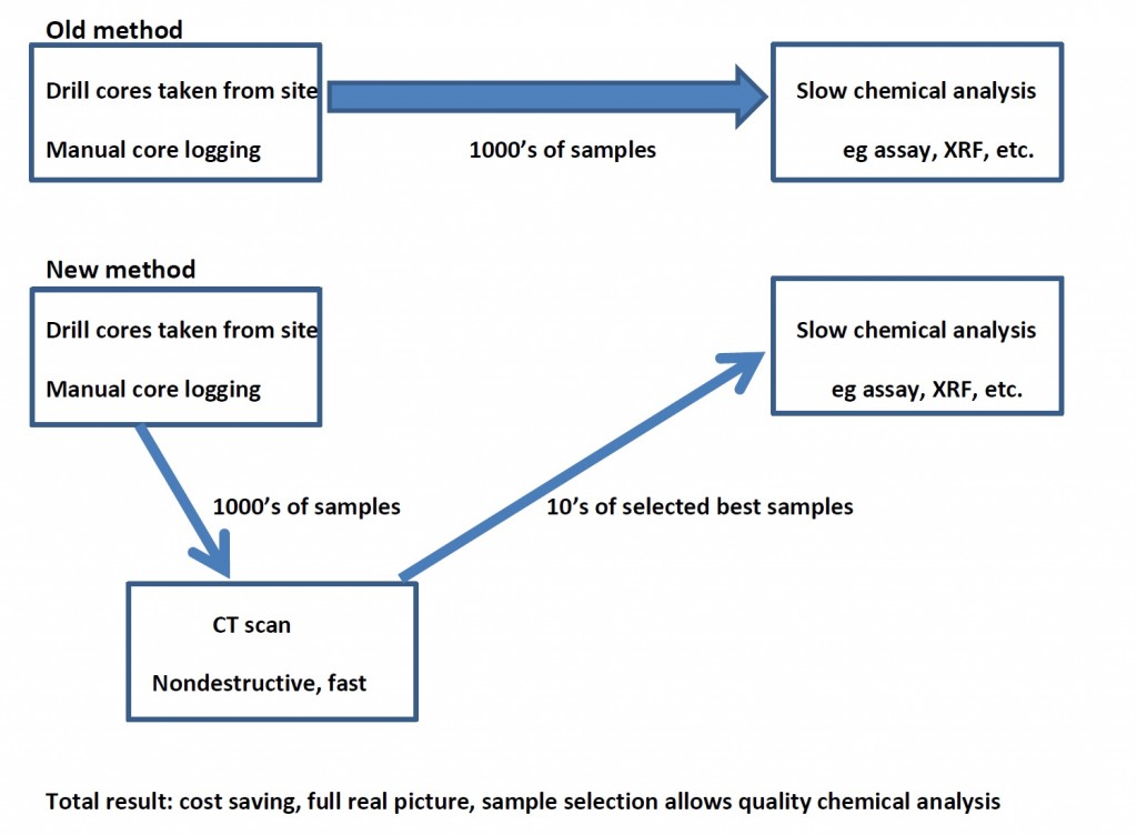
Full 3D X-ray imaging of drill cores is a fast, nondestructive method that can fit into the drill core analysis workflow, allowing the geologist to get quick images and 3D information of each drill core, non-destructively. This allows the geologist to select good samples for further analysis by traditional methods such as assay, XRF, ICPMS, etc. We demonstrate two examples here which would correspond to 1 hr per sample or 20 minutes per sample, respectively, when in large batches. This makes the method extremely cost efficient.
The workflow shows clear advantages to the geologist in improved access to information, improved quality of final information and total cost saving in the analysis workflow.
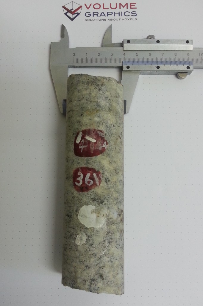
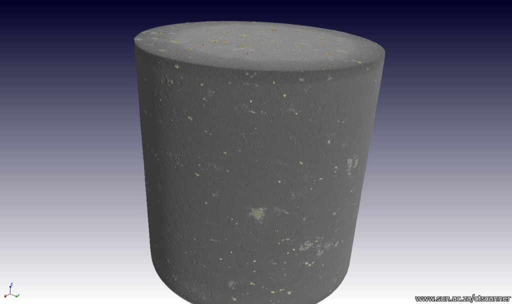
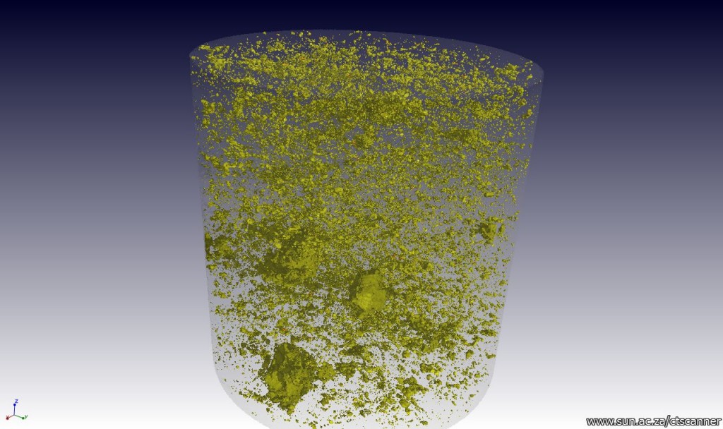
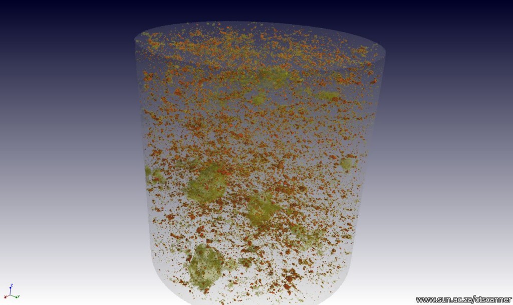
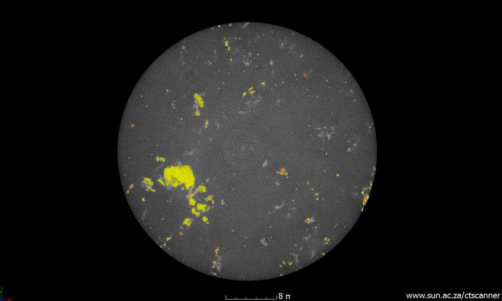
In the 1st example we present a drill core with two types of dense mineral particles at 0.1 % an 1.2%. Due to the size of the particles, a high resolution scan is required, with good scan quality, resulting in a 25 mm section imaged and analyzed in 1 hr. The photograph, its CT images and analysis results are shown below.
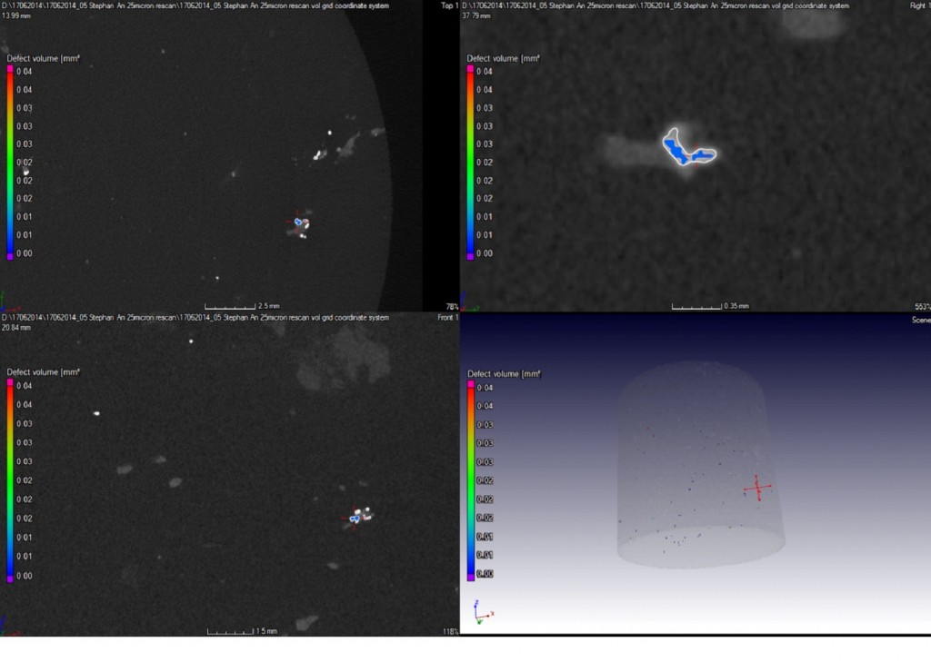
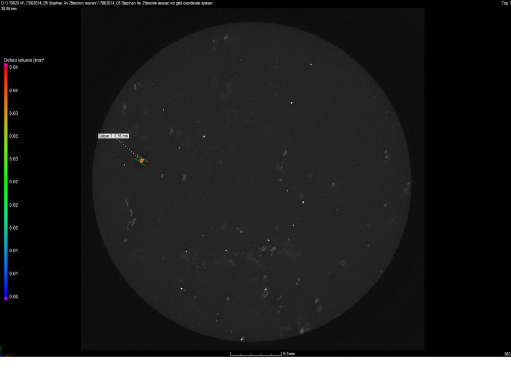
An inclusion analysis is possible on either type of inclusion, which can provide statistical information on average particle diameter, sphericity, etc. Here some of the inclusions are highlighted in an analysis.
Summary:
Non-destructive, fast CT scans provide full picture of your cores or geo-samples in a few hours
Scan times vary depending on sample type, this example was 1 hr including analysis
High quality full 3D advanced analysis capabilities adapted to geosciences
CT does not replace any technology, it fits in the workflow of geo-analysis by fast nondestructuve analysis as shown below, resulting in better total analysis, quick answers and potential cost saving in total.
Check this link for a great video of this example: https://www.youtube.com/watch?v=NEAlv_E4nOo
Analysis performed in VGStudioMax 2.2


 Follow