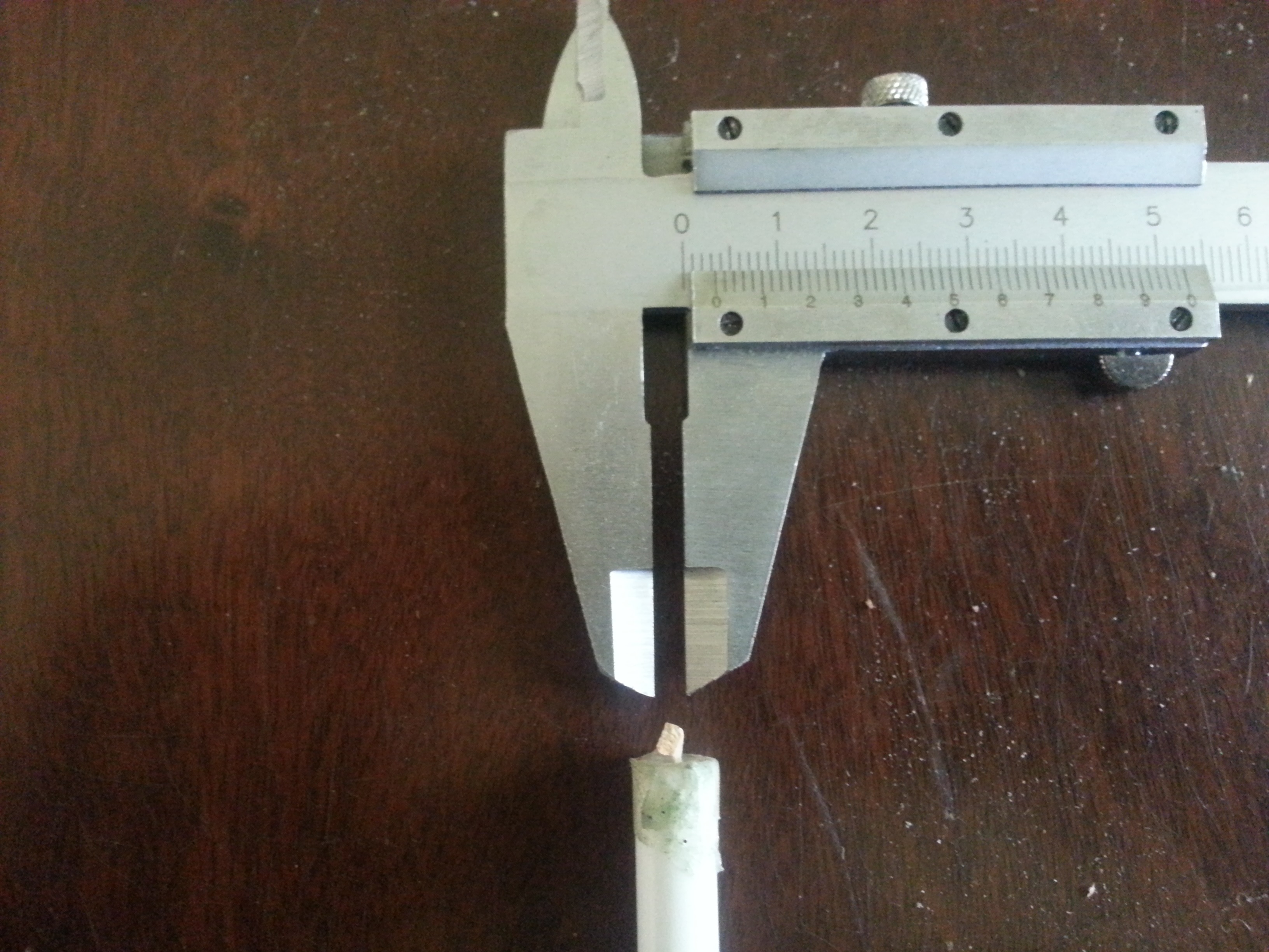
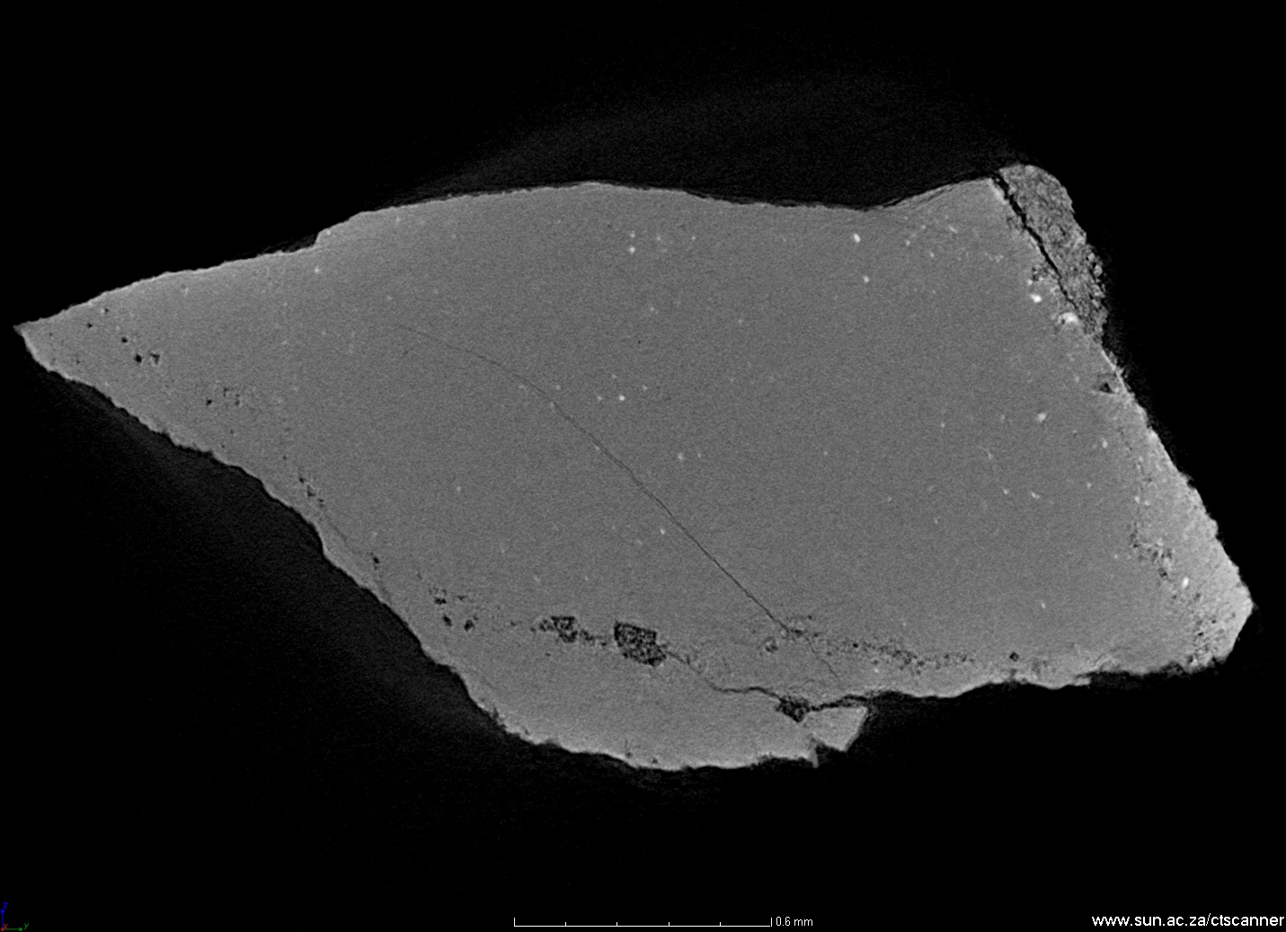
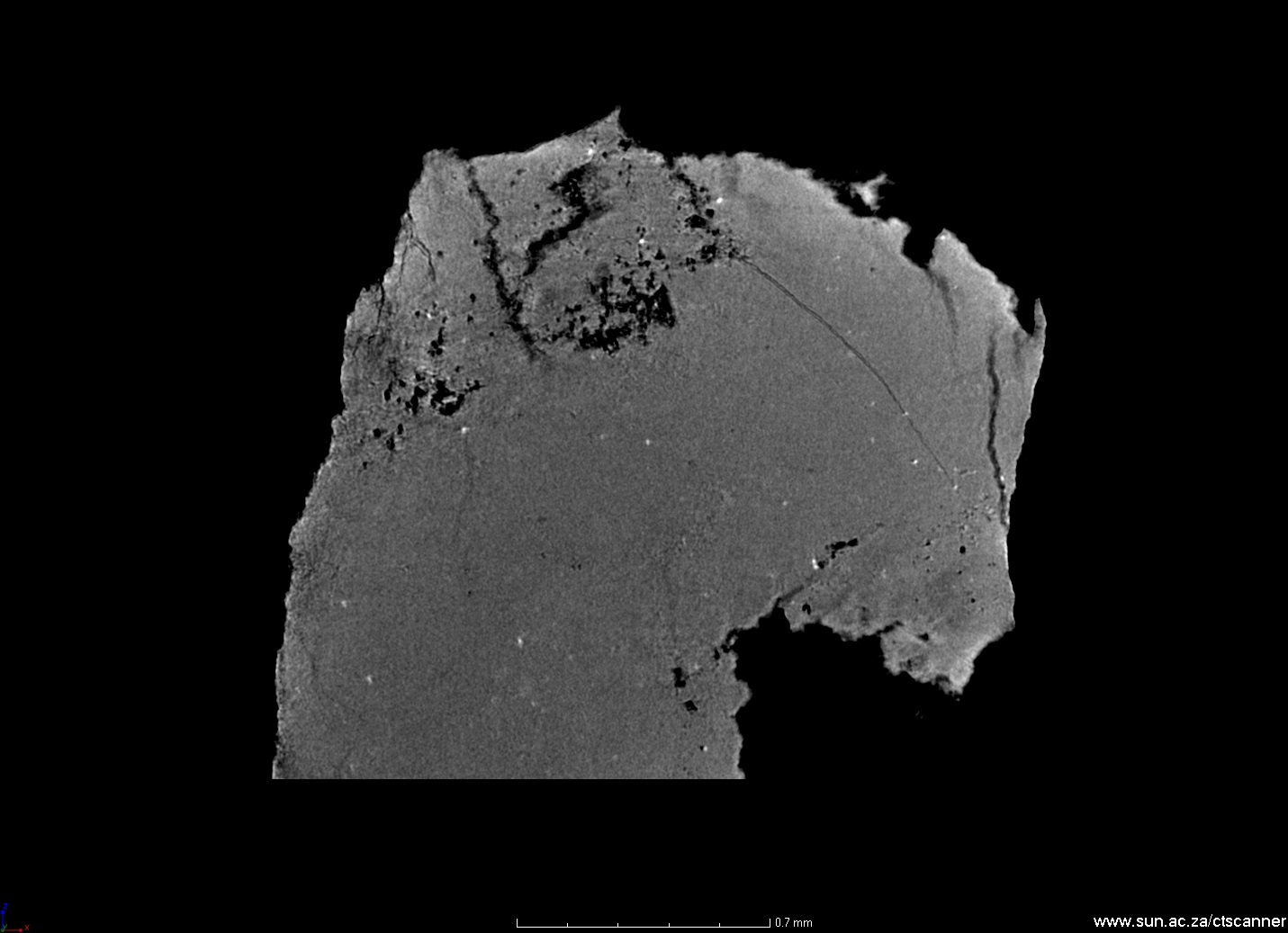
This example showcases the capability for X-ray microscopy. In some small samples, it is very difficult to physically cut and image the sample using SEM or optical microscopy, especially when looking for porosity. In this example a rock fragment is imaged, showing very high resolution detail such as porosity and inclusions. This was done at 2 micron resolution and the size of the physical sample is shown in the photo. Similar scans will be possible with the new submicron CT, allowing 500 nm resolution.
 Follow