Category: Rocks

Oct 30

Investigating microwave induced cracks

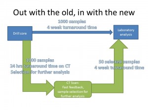
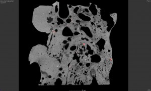
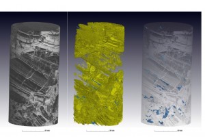
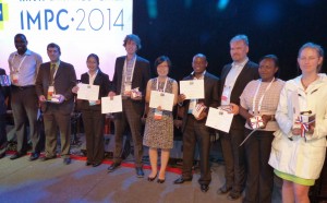
Edson Charikinya won the best young author award at the International Mineral Processing Congress in Chile, for his paper: “Use of X-ray computed tomography to investigate microwave induced cracks in sphalerite ore particles”. Edson is a final year PhD student from Stellenbosch University Chemical Engineering. His award winning presentation and a corresponding poster from another …

Continue reading

Aug 29

CT geoscience example videos

Please see below videos from various applications of CT in geosciences, for more information on these examples see the latest posts or the August newsletter which can be found CT News 2014 August 2014. Granite drill core showing distribution of two types of dense inclusions https://www.youtube.com/watch?v=NEAlv_E4nOo Hematite (iron ore) drill core showing the type of …

Continue reading

Aug 26

Drill core CT

Full 3D X-ray imaging of drill cores is a fast, nondestructive method that can fit into the drill core analysis workflow, allowing the geologist to get quick images and 3D information of each drill core, non-destructively. This allows the geologist to select good samples for further analysis by traditional methods such as assay, XRF, ICPMS, …

Continue reading

Aug 26

Slag sample: advanced analysis

The porosity of samples can be visualized and analyzed with CT scans and automated defect analysis. In addition, new tools are available to do permeability analysis of very porous materials – that means to investigate how gas would flow through a rock, for example. This is especially important for oil & gas mining industries. We …

Continue reading

Aug 26

Iron ore drill core

Even a dense iron ore core can be scanned, in a very short time, to provide visual information of the core in 3D, and measure void volume, for example. This type of scan can cover 120 mm of length in 4 samples per hour at the quality shown here Check these videos from this example …

Continue reading

May 19

Rock porosity

This example showcases the capability for X-ray microscopy. In some small samples, it is very difficult to physically cut and image the sample using SEM or optical microscopy, especially when looking for porosity. In this example a rock fragment is imaged, showing very high resolution detail such as porosity and inclusions. This was done at …

Continue reading

Nov 14

Gold Ore Project

drill core gold