
Anton du Plessis
Author's posts
Feb 09
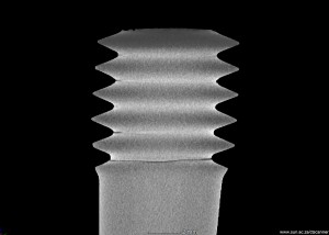
Image Quality Indicator
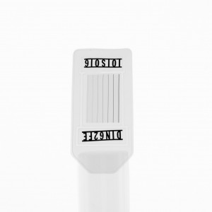
Image Quality Indicators (IQIs) are used in commercial 2D X-ray inspections to confirm detail detectability of the system. In the simplest case these are wires with well known thicknesses. It is not generally well known that 3D X-ray systems can also make 2D inspections very easily and shown below are such images of a standard …
 Follow
Jan 28
3D X-rays of Radio control airplane engine
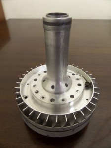
In this example a typical 3D X-ray analysis is presented of some radio-controlled airplane engine parts. Thanks to Clinton from Micton Hobbies in Somerset West, for loaning us the parts. Here are photos of the parts: The engine casing is a light metal casting, containing pores/defects. The 3D images clearly show all the defects down …
Jan 12
First print from CT data
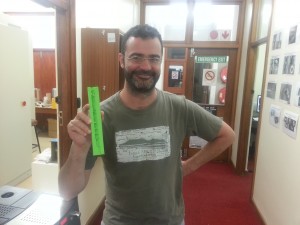
Our first 3D print from a nanoCT scan data set (blown up from 1 mm to 150 mm length) was successful! In this image the happy client is holding his 3D print of a microstructure within his material. This is a great way to visualize and demonstrate 3D information, great for presentations. When added to …
Jan 10
First 3d print
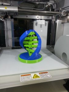
Our first 3D print is completed! As experts in 3D scanning, taking visualization to the next level with physical prints is something we’ve been looking forward to for a long time. This first print is a DNA helix – not a CT Scan data set just yet. 3D printing will be offered as an accessible …
Jan 06
Cracked Bolt
Two of the four bolts holding the engine of a 5 year old car of a well known German brand broke off, apparently after driving over a Johannesburg speedbump. The customer sent the broken bolt for a microCT scan for further inspection, with images showing the results below (cracks are black lines). Clearly, there are …
Nov 04
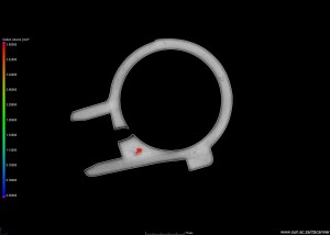
Oil pump
One part of an oil pump was scanned to demonstrate the quality of 3D information, which can be used for quality inspection or reverse engineering. See images below of slice and 3D images of internal porosity. Clearly there is one very large pore (coloured in red) which is shown in slice images also. Close-up of …
Nov 04
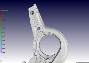
Automotive casting – water pump
This example shows a water pump, with a porosity/defect analysis performed on one region of the casting. This shows how easily casting porosity can be identified and quantified (each pore has its void volume and other parameters calculated). The analyzed region has its surface data also available for output to STL format, for reverse engineering …
Oct 30
Investigating microwave induced cracks

Edson Charikinya won the best young author award at the International Mineral Processing Congress in Chile, for his paper: “Use of X-ray computed tomography to investigate microwave induced cracks in sphalerite ore particles”. Edson is a final year PhD student from Stellenbosch University Chemical Engineering. His award winning presentation and a corresponding poster from another …
Oct 30
Biology – looking inside brittlestars
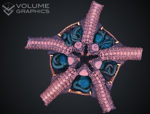
This work is from Jannes Landschoff, who won the best poster award for his poster “How many inside? A 3D Micro CT-Scan of Brooding Ophiuroids” at the European Echinoderms Colloquium, Portsmouth, UK in July. Jannes is a MSc student from UCT Biology. This work is still ongoing so more striking images will be loaded once …
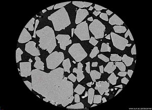
Oct 30
Particle size distribution analysis
X-rays imaging at high resolution allows visualization and quantification of any type of sample, including particles and powders. X-ray computed tomography in the micro and even nano scale is now possible, as a service facility in Stellenbosch University. Here we present the advantages of using the method for particles and powder characterization. The full 3D …