
Anton du Plessis
Author's posts
Oct 30
Particle size distribution analysis
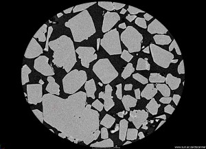
X-rays imaging at high resolution allows visualization and quantification of any type of sample, including particles and powders. X-ray computed tomography in the micro and even nano scale is now possible, as a service facility in Stellenbosch University. Here we present the advantages of using the method for particles and powder characterization. The full 3D …
 Follow
Sep 26
NanoCT of a toothpick
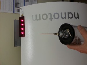
This example puts a nanoCT scan into perspective, as the 550 nm resolution allows a field of view of up to about 1 mm, allowing the tip of a toothpick for example to be analyzed in full 3D. This is why this is also called a 3D X-ray microscope. See below some images and videos …
Sep 25
Swimming pool analysis
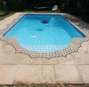
No we cannot CT scan your whole swimming pool, not non-destructively anyway. What we can do is take a sample of fibreglass from where the weir is cut out, and analyze that section. In this example, two samples of different fibreglass pool manufacturing methods are compared. These are the chopper gun application (sample 1) and …
Sep 25
Damaged High Voltage Insulator
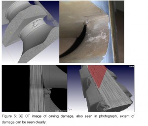
In this proof of concept, a damaged HV insulator used in power line transmission was scanned to see if internal damage can be assessed positively. The idea is to scan these objects preventatively before serious failure. Some images show the damage subsurface and the clarity of routine-type 3D X-ray inspection.
Sep 25
Carbon fibre with paint coating
In this example, thanks to one of our clients, we can share some images of a carbon fibre material with a painted coating. We show how the coating thickness can be assessed with an automatic “wall thickness analysis” across the field of view, indicating variations in thickness by colour changes. A fibre orientation analysis shows …
Aug 29
CT geoscience example videos
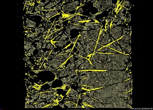
Please see below videos from various applications of CT in geosciences, for more information on these examples see the latest posts or the August newsletter which can be found CT News 2014 August 2014. Granite drill core showing distribution of two types of dense inclusions https://www.youtube.com/watch?v=NEAlv_E4nOo Hematite (iron ore) drill core showing the type of …
Aug 26
Drill core CT
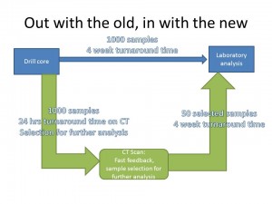
Full 3D X-ray imaging of drill cores is a fast, nondestructive method that can fit into the drill core analysis workflow, allowing the geologist to get quick images and 3D information of each drill core, non-destructively. This allows the geologist to select good samples for further analysis by traditional methods such as assay, XRF, ICPMS, …
Aug 26
Slag sample: advanced analysis
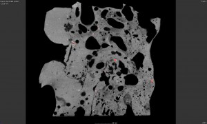
The porosity of samples can be visualized and analyzed with CT scans and automated defect analysis. In addition, new tools are available to do permeability analysis of very porous materials – that means to investigate how gas would flow through a rock, for example. This is especially important for oil & gas mining industries. We …
Aug 26
Iron ore drill core
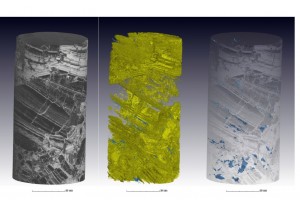
Even a dense iron ore core can be scanned, in a very short time, to provide visual information of the core in 3D, and measure void volume, for example. This type of scan can cover 120 mm of length in 4 samples per hour at the quality shown here Check these videos from this example …
Aug 26
NanoCT launch and NIR training
The Stellenbosch University CT Scanner Facility is expanding its capabilities into the nanoscale. Please join us in launching our new instrument and come meet some other users of this diverse and exciting facility. The invitation is attached here, all interested parties are welcome, also your partners, but please RSVP: nanoCT invite Thanks to the NRF, …