
Anton du Plessis
Author's posts
Mar 24
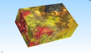
Scanning bones
In a recent academic project, a batch of scans was done on bovine bone samples at 80 micron resolution. This works forms part of a collaborative study between UCT (RSA) and ENSAM (France) aimed at understanding mechanical strength of bone so as to improve the design of protective clothing and structure. Due to the size …
 Follow
Mar 03
3D nondestructive imaging of garlic flakes
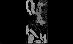
This proof of concept study shows how non-destructive imaging can be used to visualize cellular structures and thereby identify different materials, in this case garlic and some other biological material related to garlic. In this slice image, the green arrow indicates garlic with its unique cell structure and density (grey value), while the red arrows …
Feb 10
CT and MRI scanning
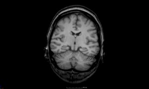
As a complementary 3D imaging tool for our clients, we offer the ability to do experiments using MRI (magnetic resonance imaging) scans in collaboration with our colleagues at the Brain Imaging Centre. MRI is complementary to CT and allows the visualization of especially water-rich materials such as biological tissue: live animals, meat, fruits, plants, etc. …
Feb 07
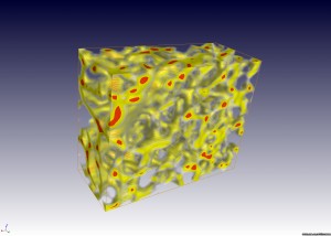
Digital Volume Correlation
In a unique and powerful collaboration between our facility and the Mechanical Engineering Dept at the University, we are offering a new service: Digital Volume Correlation, using the LAVision commercial software package. The software calculates changes occurring in a sample in full 3D, making it possible to quantify and visualize changes in 3D using colour …
Jan 10
Anatomy of a shrimp
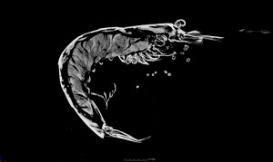
There are many applications of microCT in scanning small insects and small animals, for positive identification of their species and 3D visualization of internal and small structures, where traditional microscopy sometimes fails to produce good images. This is an example of a shrimp, with 3D views, a slice view and some measurements of limb lengths, …
Jan 10
Pharmaceuticals
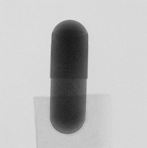
Have you ever wondered how a pill looks like on the inside? In this example, a headache capsule from a leading pharmaceuticals company was scanned to demonstrate the internal packing and density variations. This kind of inspection could be useful to optimize production, limit the inclusion of metallic or other dense particles and ensure even distribution of …
Jan 10
Light bulb
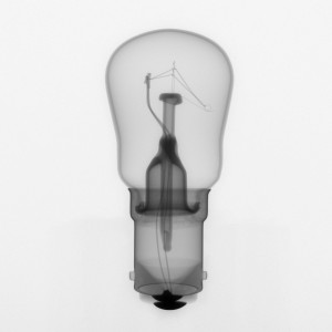
A simple example of the X-ray imaging capabilities of this technology is with a normal filament-type light bulb. A digital X-ray image (obtained in a few minutes) is shown, providing useful information on the location of different materials and providing an inside view, though in 2D only. This type of imaging is a standard NDT …
Jan 10
Wine volume

A microCT scanner is a precision dimensional measurement device, making it possible for example to measure the volume of wine inside a closed bottle of 1963 South African sweet wine (Port). The images illustrate the bottle (semi-transparent), the lead neck cover (green), the wine (dark red) and the air in the neck of the …
Nov 25
Casting CT inspection
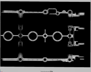
Metal castings are usually inspected by normal X-ray images but the enhanced contrast of X-ray CT allows clear identification of the location, type and potential effects of defects without any doubt. The example here was a vehicle Cam cover, the images illustrate the largest defects, and for potential clients interested in this inspection service we can provide for this …
Nov 25
Characterizing sweet potato powders
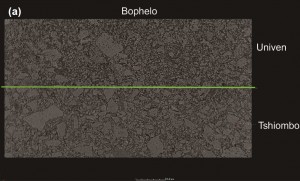
In this Food Science research project from the University of Venda, sweet potato powders were studied as grown at different sites, in the images below from the Bophelo cultivar, the powder from the Univen site contained more smaller powder particles. Further work is expected and the powder characterization in 3D, as well as density differences …