The Hans Merensky Chair in Measuring and Modelling Eucalypt Growth & Wood Formation
Understanding growth in the world's most widely planted hardwoods
Update on Letitia’s CT scanning experiment
Post authored by Dr Letitia Schoeman
In an effort to explore X-ray computed tomography (CT) as a non-destructive approach to visualise and quantify the developmental processes in differentiating xylem in eucalypts, an experiment was developed to investigate how the water availability preceding a severe drought affects cambium development in Eucalyptus grandis clones.
The development of methods that can non-invasively visualise plant microstructure and characterise changes that occur during growth and development is of paramount importance. In Eucalyptus, cambium development involves multiple complex interactions and to describe and understand this development, a non-destructive method for visualising and quantifying developmental processes is potentially very useful.
In this study, which were performed in a growth chamber (Figure 1) under controlled conditions, the development of the cambium in living Eucalyptus grandis clones was tracked for four weeks. To determine the response to repeated drought stress, two irrigation treatments (well-watered and droughted) were applied. These respective treatments were followed-up by a severe drought and a subsequent re-watering phase to monitor the recovery.
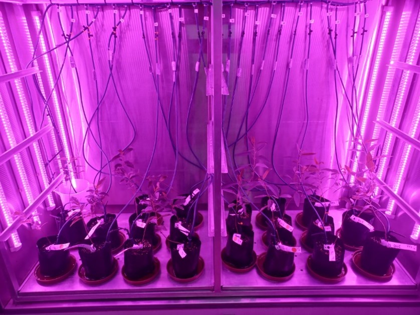
Figure 1: Growth chamber containing the Eucalyptus grandis clones, where each sample has its own watering pipeline connected to it for the respective well-watered and drought treatments.
All the samples were scanned on a weekly basis making use of non-destructive X-ray micro-CT (Figure 2). Micro-CT systems have a limited resolution which is not sufficient for analysing the cellular structure. Thus, after the last week’s micro-CT scans, high-resolution nano-CT (Figure 3) was also performed to gain more detailed cellular information of the cambium in the same plants. For the micro-CT scans the whole sample was scanned at parameters of 90 kV and 170 μA, which achieved suitable image quality with a voxel size of 9.5 μm. For the nano-CT scans a sub-section of the same ROI of the stem was scanned at parameters of 80 kV and 180 μA, attaining a resolution of 1.5 micron.
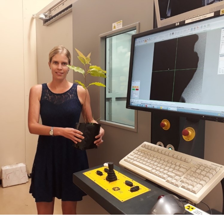
Figure 2: The General Electric V|Tome|X L240 X-ray micro-CT system used to scan the whole Eucalyptus grandis clone.
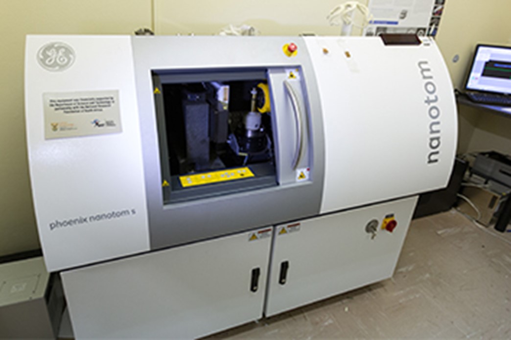
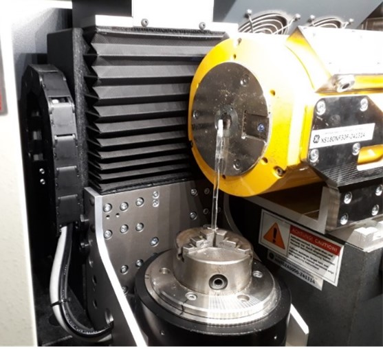
Figure 3: The General Electric Nanotom S nano-CT system used to scan a sub-section of the plant stem. Samples smaller than 10 mm in diameter works best on this system.
Letitia will be spending the next couple of months analysing her data at the high-performance data analysis facility at the CAF CT-scanner unit (Figure 4). During image analysis specialised VGStudio Max software is used to perform 2D and 3D characterisation to obtain quantitative results such as dimensional and volumetric measurements.
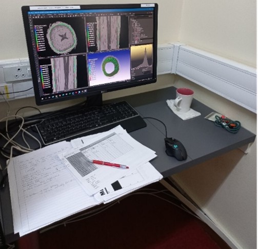
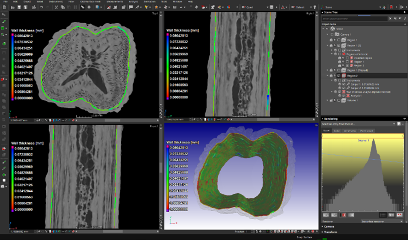
Figure 4: The high-performance data analysis facility where workstations can be booked to perform analyses. In the example, the wall thickness analysis module was applied to characterise the cambium tissue, providing a 3D color-coded distribution that localize areas with varying thicknesses.
This study will shed some light on whether previous exposure to limited water conditions may provide increased protection during future water deficit or droughts and how the cambium development changed during these conditions. We are excited to see what the results look like!