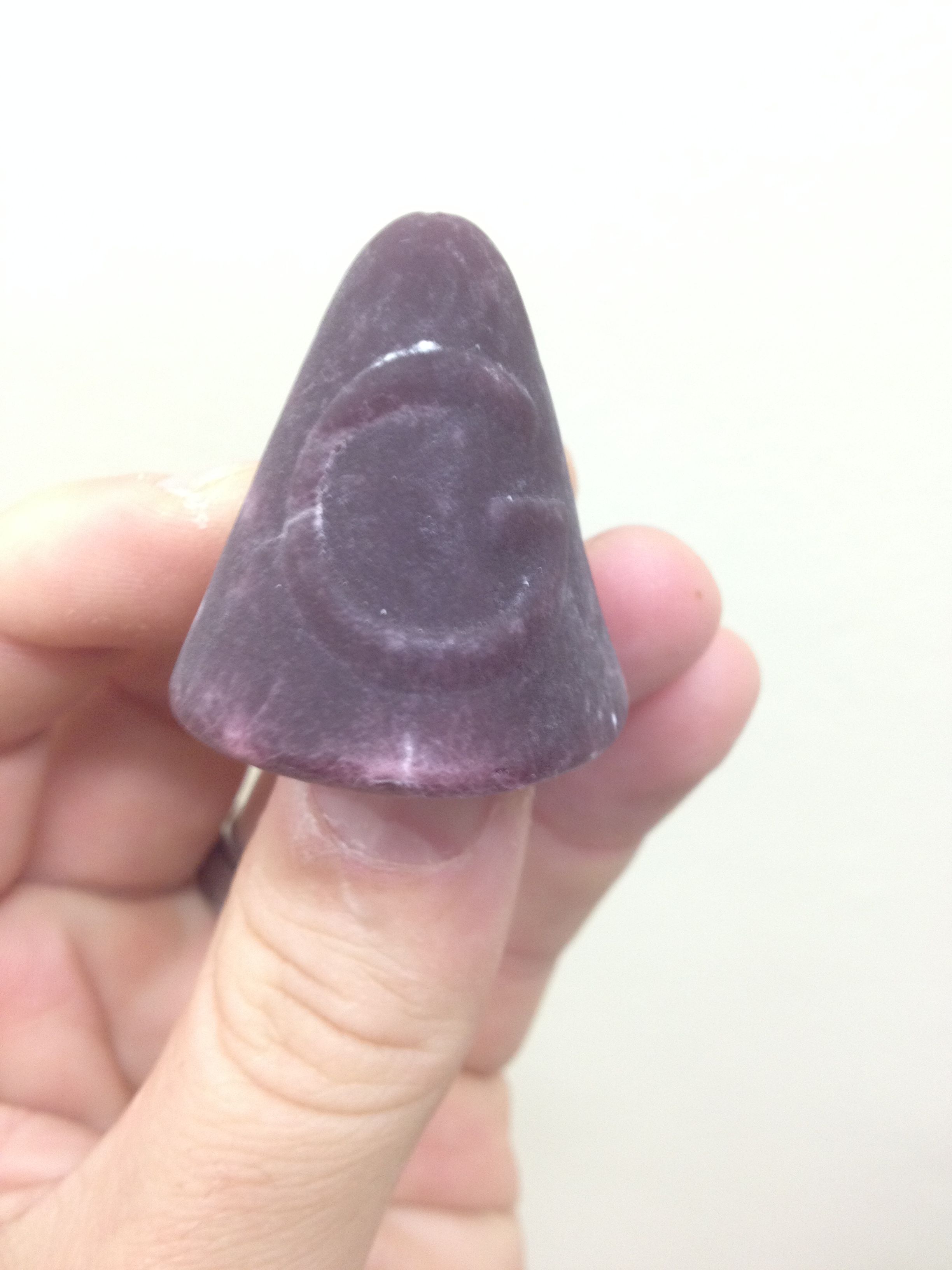
A Ghent nose is a sweet originating from Ghent, Belgium. Since we visited Ghent last month, we decided to scan and analyze a Ghent nose as an example. This example shows how 3D X-ray imaging can be used to visualize internal features of food products, in this case showing clearly some large air bubbles / pores. The colour is added to show the volume of the air bubbles. Besides these images, two videos show more information and below that additional images show 2D X-rays and 3D CT slice images.
Enjoy the sweet treat!
Videos corresponding to the story below are found here:
crop nose to show big air bubbles
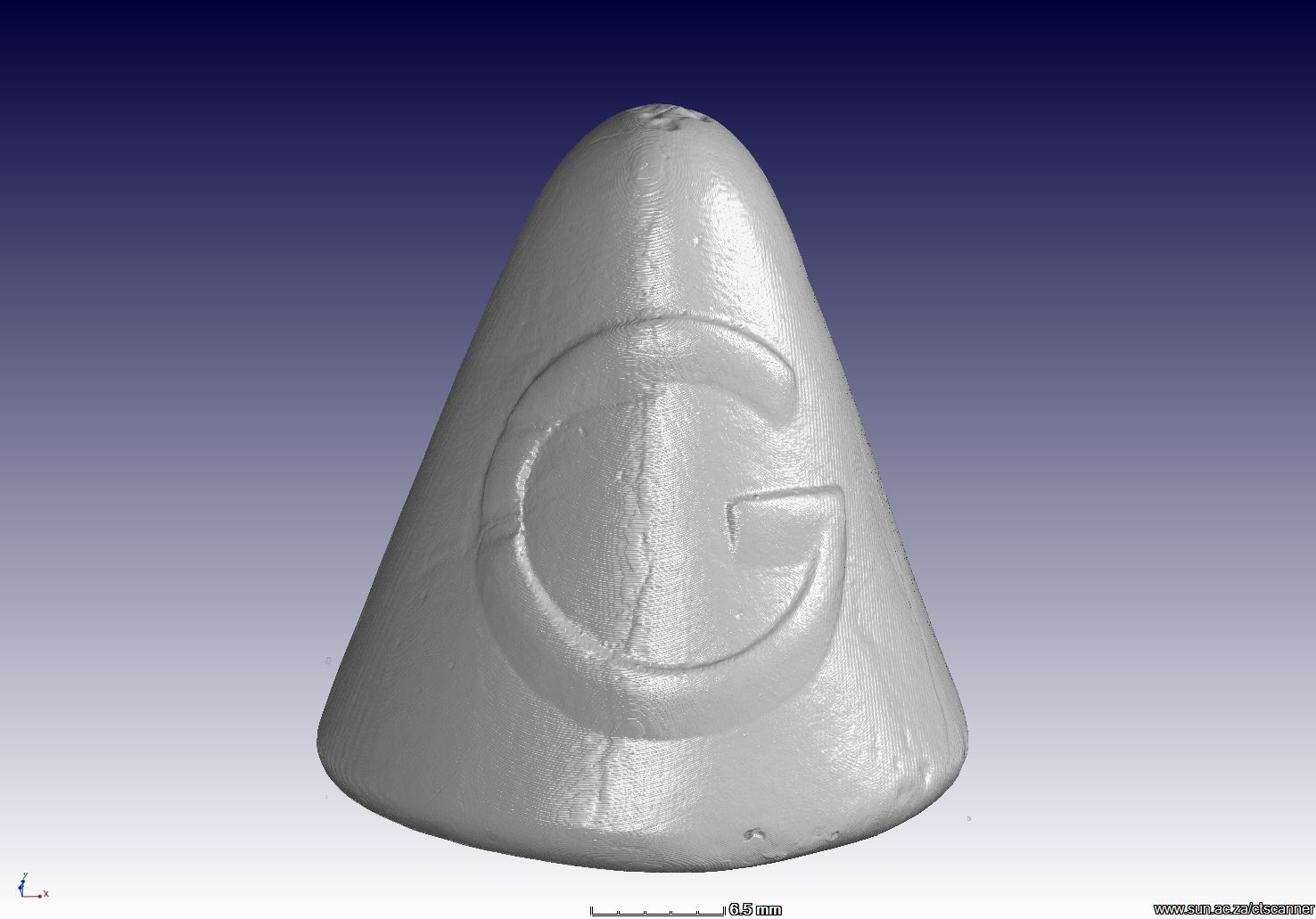
First a 3D surface view from the CT scan data, showing the G which stands for Ghent:
The a defect analysis highlights the air bubbles / pores in colour coding according to size and a crop window cuts open the view to reveal the internal features:

A semi-transparent view shows wth shadows the location of the internal semi-liquid compared to the crunchy outer layer, in addition to the internal air bubbles:

A full transparency shows the air bubbles clearly: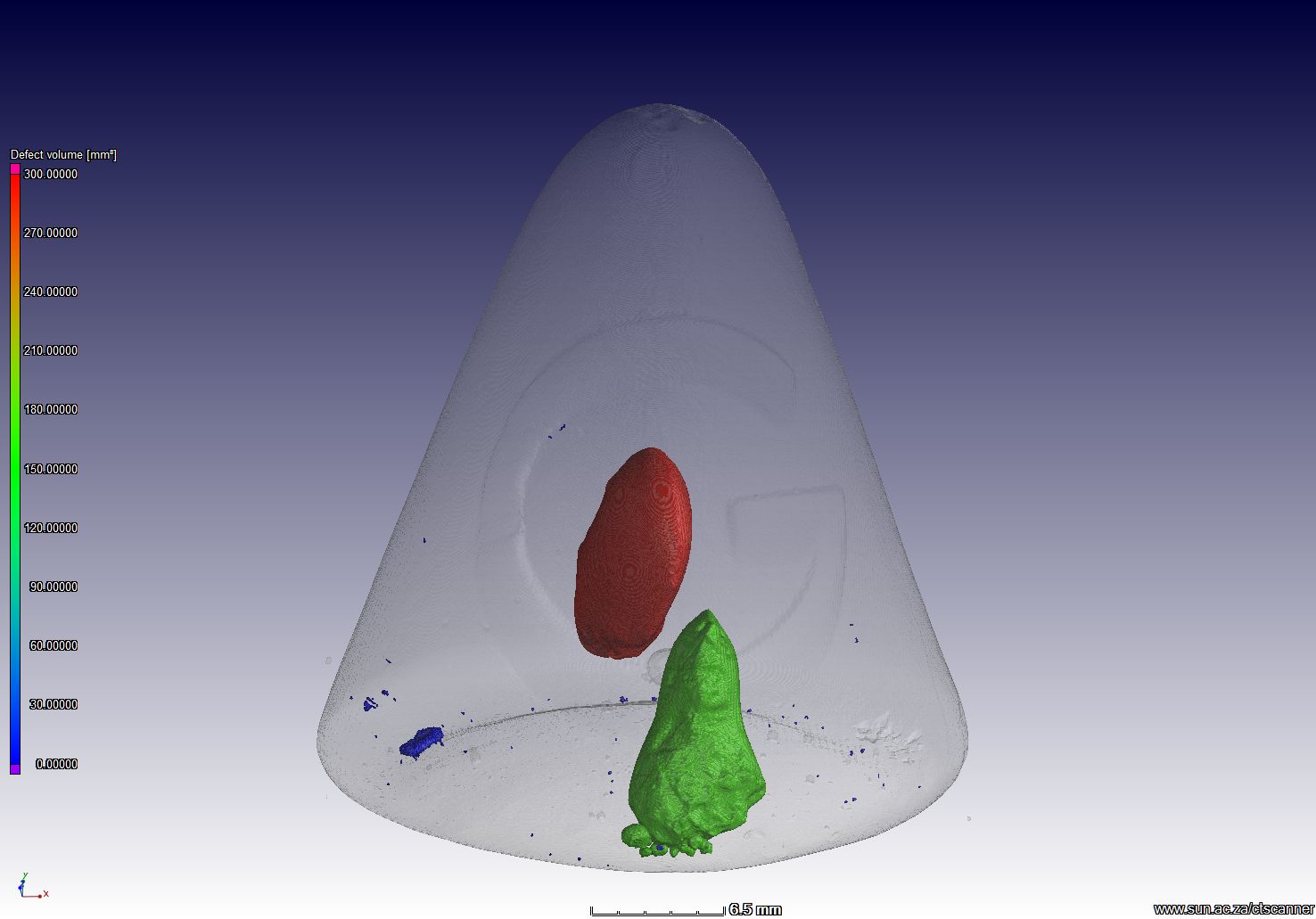
A wall thickness analysis applied to the crunchy outside layer, shows the layer is thickest on the top (red):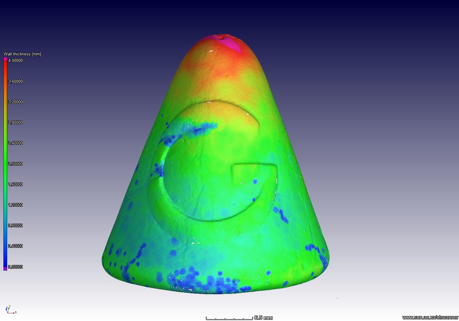
A surface determination with triangulation is used to output a STL file for example for 3D printing, here the triangle mesh from CT scan data is shown in blue with more images below showing more detail of the mesh: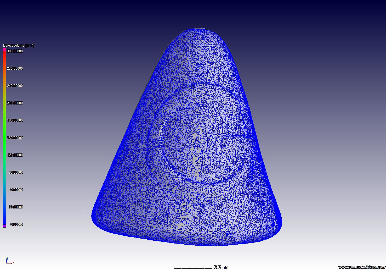


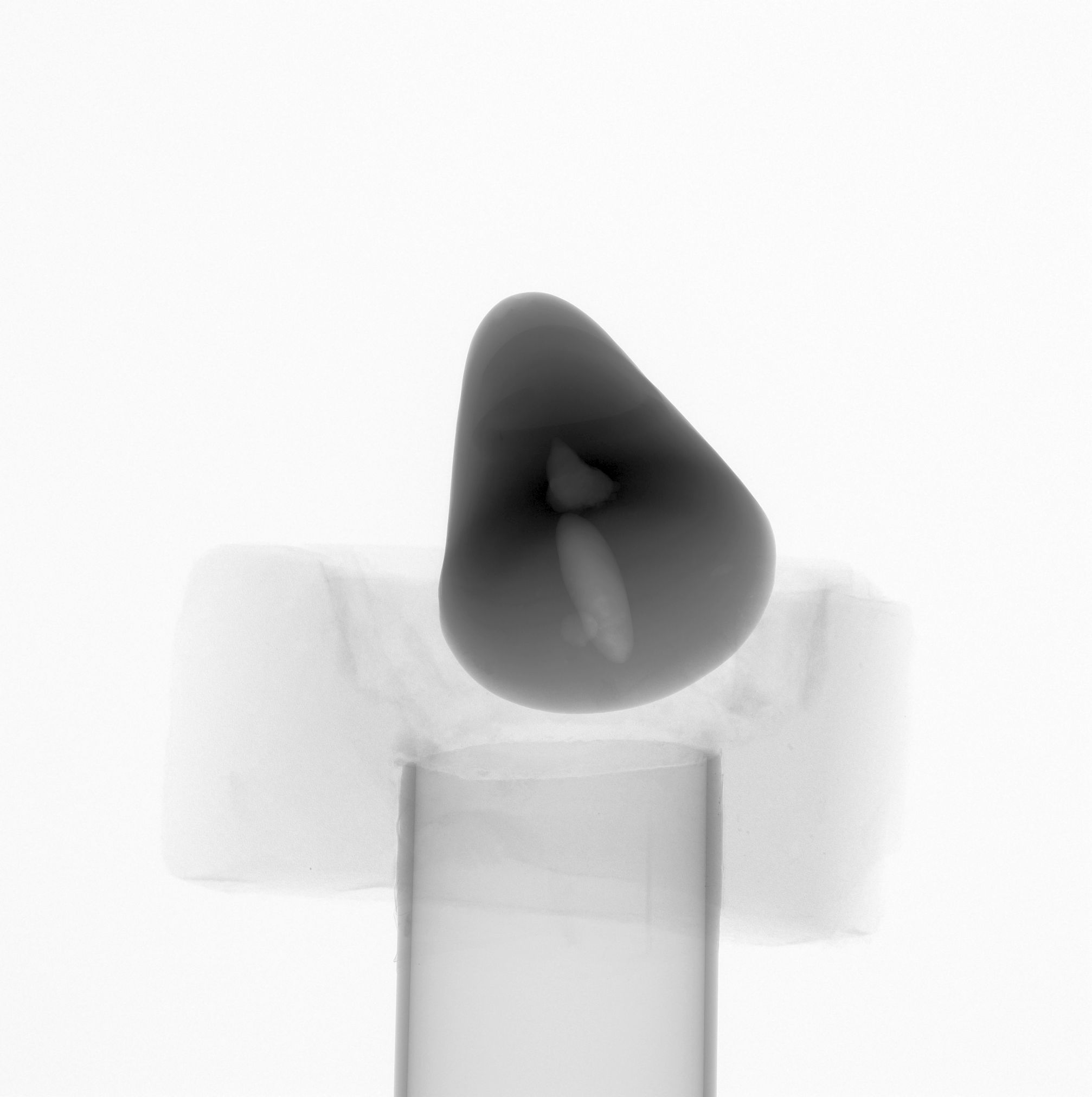
Here is an X-ray image of the sample in the scan:
This is a CT slice image showing the contrast obtained on the pore / bubble space: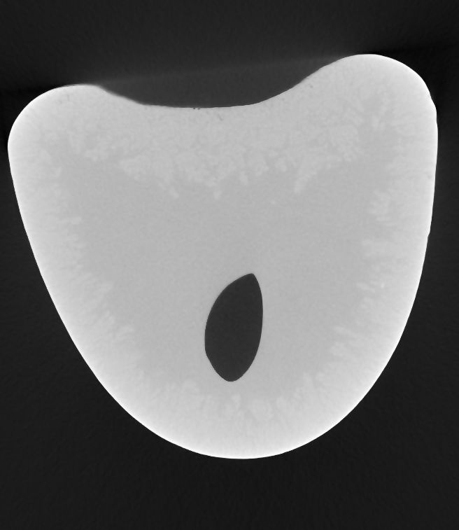
Analysis performed by Volume Graphics VGStudioMax 2.2
 Follow